Cholesteatoma & its Treatment
Cholesteatoma
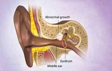
A cholesteatoma is an abnormal, noncancerous skin growth that can develop in the middle ear, the cavity behind the eardrum where we find the three small bones that form the ossicular chain. It is most commonly caused by repeated middle ear infections but can also result from a birth defect.

Why does a cholesteatoma develop?
We all have skin inside our ear canal. It is meant to be there and is a normal part of our ear. But with a cholesteatoma the skin right next to the eardrum, deep in the ear, gets sucked in gradually to where it shouldn't be. This can happen when the eardrum becomes retracted due to poor ventilation from the Eustachian tubes. A retraction pocket can develop and the skin then forms a tiny pearl, or ball, that keeps burrowing its way deeper into the ear over many months. It damages the delicate bones inside the middle ear - the bit that is responsible for hearing. At this point it becomes painful.
If left untreated it will push further and further inside the ear, through the inner ear and possibly even next to the brain.
There are two types of cholesteatoma:
Congenital cholesteatoma: is a problem that in theory happens from birth. For some reason, even though the eardrum is normal, tiny skin cells get sucked into the middle ear, blocking the Eustachian tube. This then causes long-term fluid in the middle ear (which is usually free of fluid) and can cause hearing loss. This becomes apparent between the ages of 6 months to 5 years when the child's hearing doesn't develop properly. This is a very rare condition and the cause isn't fully known.
Acquired cholesteatoma: develops later, usually in adults between 30 and 50 years old. Again, the cause isn't fully known. Sometimes a cholesteatoma in an adult can happen from having a grommet - a tiny tube that is put through the eardrum as a treatment for middle ear problems - as a child
The true occurrence rate of cholesteatoma is not known. About 1 in 1,000 people with ear problems referred to ENT clinics have cholesteatoma. It has also been suggested that there is about 1 case per 10,000 population. Most cases are of the acquired type.
Symptoms of Cholesteatoma
The symptoms associated with a cholesteatoma typically start out mild. They become more severe as the cyst grows larger and begins to cause problems within your ear.
Initially, the affected ear may drain a foul-smelling fluid. As the cyst grows, it will begin to create a sense of pressure in your ear, which may cause some discomfort. You might also feel an aching pain and fullness in or behind your ear. Sometimes facial numbness is present.
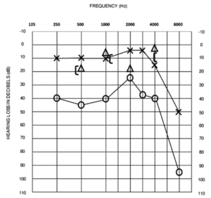
The pressure of the growing cyst may also cause hearing loss in the affected ear so audiological evaluation is an important step to diagnosis.
Diagnosis
Audiological assessment.
CT scan of the middle ear provides images of the cyst.
Examination under microscope to confirm findings.
Magnetic resonance imaging (MRI) scan of the brain is performed when the brain is suspected to have been affected.
Treatment
Although surgery is rarely urgent, once a cholesteatoma is found, surgical treatment is the only choice. Surgery usually involves a mastoidectomy to remove the disease from the bone, and tympanoplasty to repair the eardrum. The exact type of operation is determined by the stage of the disease at the time of surgery
Mastoidectomy: When an infection or cholesteatoma has grown into the mastoid (the bone behind your ear), we will open the bone to remove the disease. Sometimes this requires removing all the bony walls within your ear to create an open cavity. This process is called an "open cavity," "canal wall down," or a "radical mastoidectomy." Sometimes the bony partitions can be preserved, resulting in a canal wall up”, or “closed cavity” mastoidectomy.
Tympanoplasty: This refers to repair of the eardrum and hearing mechanism. The eardrum is repaired with a graft of cartilage or fascia (the lining of the muscle behind your ear). Usually this type of repair is successful in closing holes in the eardrum permanently. If the small bones involved in hearing are damaged, we will try to repair them with natural bone or cartilage, or synthetic prostheses made of bone mineral. We use natural bone whenever possible.
Cholesteatoma surgery, which is delicate surgery performed under a microscope, usually takes 2 to 3 hours, and patients may go home the same day. It is very important to remove the disease completely, or it may grow back. The rate of re-growth is higher in children than adults. Because of the aggressive nature of cholesteatoma, we ask patients come in regularly for careful follow-up. Sometimes a second operation will be necessary.

